
MRI Scanners

G-scan Brio — nowy rezonans magnetyczny w obciążeniu
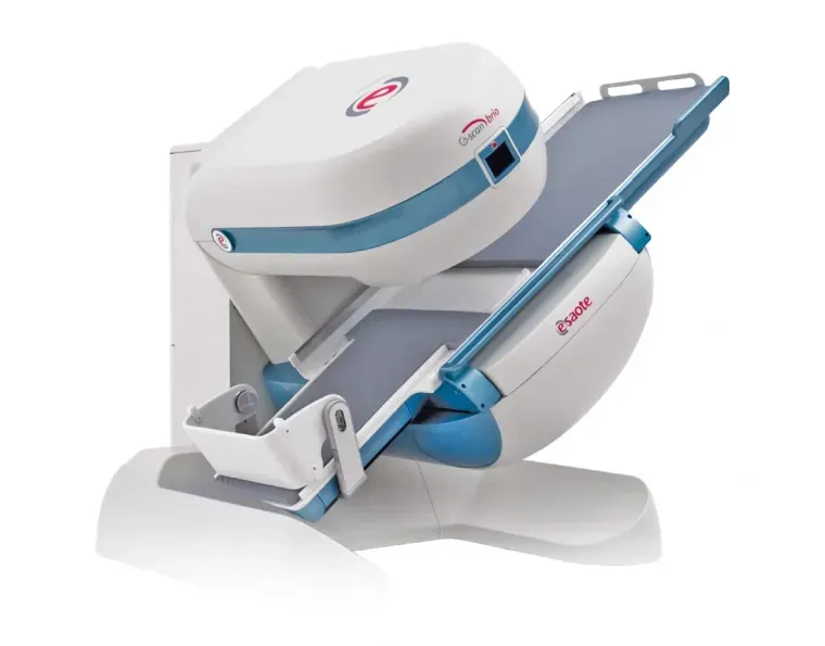
G-scan Brio is an open Esaote magnetic resonance imaging system for diagnostics of the locomotor system, enabling examination of the patient under natural load.
Key features
REZONANS W JEDNYM POMIESZCZENIU
23 m². Minimalna powierzchnia instalacji, system plug and play.
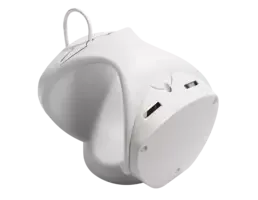
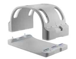
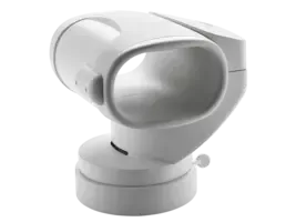
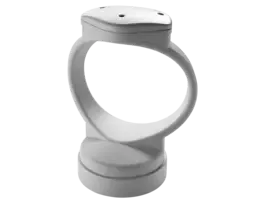
DEDYKOWANE CEWKI WIELOZADANIOWE
12x. Zaprojektowane do badań kręgosłupa i stawów.
DEDYKOWANE SEKWENCJE
2x. Możliwość połączenia badania w obciążeniu z obrazowaniem dynamicznym.
EKOLOGICZNY REZONANS
<3 kW. Niskie zużycie energii.
G-scan — open MRI for weight-bearing examinations
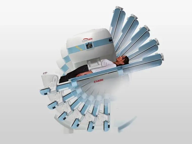
G-scan Brio is a dedicated Esaote magnetic resonance imaging system for musculoskeletal diagnostics, which allows the patient to be examined in both the supine and standing positions — under natural load. It is a solution for diagnostic laboratories, orthopedics, and sports medicine, for which it is important to reveal pathologies that intensify under the influence of gravity.
Application
A system for MRI examinations in the patient's supine and standing positions — under natural load.
Which examinations is it for
Weight-bearing examinations, all upper and lower limbs, hip, spine, head.
Cost-effectiveness of purchase
Very fast return on investment. Low fixed costs — the cost of an examination consists only of energy costs. Single-phase 230V power supply. Low installation costs. No fixed costs related to cooling the device — it does not require helium cooling.
First commercial installation in Poland
G-scan Brio is an innovative MRI system for examining the patient in the supine and standing positions
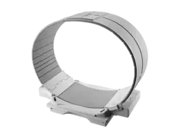
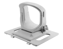
The G-scan Brio magnetic resonance imaging system provides solutions in MRI diagnostics that increase diagnostic accuracy and the accuracy of diagnosis in musculoskeletal examinations. Many years of experience have resulted in the development of an MRI system enabling examinations to be performed not only in the supine but also in the standing position — under natural load. A wide selection of dedicated coils, designed specifically to fit the examined body parts, allows all musculoskeletal examinations to be performed, and a dedicated head coil is optionally available.
The solution of changing the patient's position is particularly important for the effective diagnosis of pathologies whose image may change due to posture and body position, such as lower back pain.
Why weight-bearing MRI
Many MSK pathologies become apparent or are more clearly visible in the weight-bearing position:
- Spinal stability / instability
- Degenerative pathologies in the spine
- Osteoarticular pathologies (sports medicine)
- Pathologies of osteoarticular lesions
- Meniscus and patella lesions
Video materials about the G-scan
General characteristics
- A permanent magnet weighing about 8 tons.
- A unique magnet rotation mechanism — the magnet can be rotated from the horizontal to the vertical position, enabling imaging of physiological changes under the influence of gravity. Imaging can be performed in any position within the range from 0° to 90°. The smoothness of the table rotation is provided by a special hydraulic mechanism.
- A special, lightweight Faraday cage made of innovative materials. Thanks to its structure, it uses the existing lighting and air conditioning, reducing installation costs. Thanks to its low weight, it does not require additional construction adaptation.
- The minimum room area required for installation is 23 m2. The system only requires maintaining a constant temperature in the room.
- Low energy consumption (less than 3kW).
- A simple and intuitive operator console guarantees optimal ergonomics and minimal installation space.
- An intuitive user interface — it allows a new user to quickly start scanning, while an experienced operator will appreciate the full range of imaging sequences and the ability to program them.
- Patient positioning with real-time preview on the control console as well as on the screen built into the magnet.
- A complete set of pre-programmed protocols for greater user convenience
- Self-centering high-sensitivity coils with automatic recognition of coil filling.
- A patient bed that provides comfort as well as easy and fast positioning. Additionally, a set of dedicated cushions for better patient positioning
- No claustrophobia effect.
- The ability to image obese patients.
- The device housing is designed to increase patient comfort and ease of positioning.
- Easy assumption of the diagnostic position. Unique software for segmenting the spine structure
Easy patient positioning
Patient positioning with real-time preview on the control console and on the screen built into the magnet, no claustrophobia effect, and the ability to image obese patients.
High image quality and short scan time
Standard throughput of 2 patients / hour with preparatory procedures, fast 12-minute protocols (e.g., for knees).
Low examination cost
Only energy costs (as much as an average electric kettle).
Low room adaptation costs
The external Faraday cage and single-phase power supply do not require power modifications, as is the case with high-field MRI.
Check the differences
Dedicated G-scan magnetic resonance imaging vs. high-field magnetic resonance imaging
- Single-phase power supply
- No need to interfere with the existing installation
- Does not require power modifications, as is the case with high-field MRI
- Low examination cost — only energy costs (as much as an average electric kettle)
- Low service costs — only calibration costs
- Low room adaptation costs — external Faraday cage
- Max. power draw 2.4k VA, which is about 2000W + the power draw of the air conditioner
- The need to create a separate 3-phase power supply with 100A protection
- The need to replace (replenish) the cooling agent
- An electromagnet at specified time intervals — high costs
- High service costs
- High room adaptation costs (the need to build a Faraday cage and specialized air conditioning during room adaptation)
eXP Technology
Check the technologies found in the G-scan
- True Motion — enables real-time imaging of moving joints. The entire sequence can be saved in the popular .avi format, so the video can be played on almost any device.
- MAR Metal Artifact Reduction — software enabling the reduction of image artifacts originating from metal implants.
- 3D Viewer — an advanced viewer for 3D images created through acquisition using 3D isotropic sequences.
- 2D/3D Speed Up — software that, using mathematical algorithms, allows the acquisition and image reconstruction time to be reduced by up to 30% in 2D mode and 50% in 3D mode compared with the standard imaging mode.
- Isotropic 3D — advanced imaging of three-dimensional isotropic sequences that makes it possible to combine Speed Up technology, allowing fast acquisition of high-resolution 3D images, with advanced 3D image reconstruction techniques — MPR. In a single acquisition, we obtain the highest-quality image in all three planes.
- 3D Sharc — a three-dimensional sequence dedicated to high-resolution imaging of cartilage tissue.
Ask about an offer and purchase options for the G-scan
We will be glad to prepare an offer tailored to your facility and to advise on installation and service. We invite you to contact us.
Clinical images

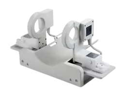
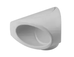
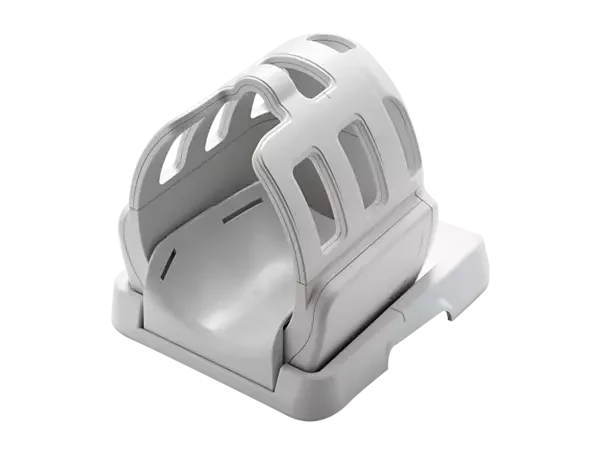
Coils
Customer care
Installation and training
We install the system and train your staff on the examinations you actually perform.
Our own service department
730,000 km a year to clients across Poland. Inspections, repairs and original parts.
Application support
Contact with an application specialist for as long as you use the system.
Product details
Key technologies
| Weight-Bearing MRI | Magnetic resonance imaging in the weight-bearing (standing) position, revealing pathologies that are emphasized under gravity. |
|---|---|
| Open and Tilting Design | Open and tilting design allowing patients to be examined from the vertical standing position to the traditional supine position. |
| Dynamic Imaging | Possibility to combine the weight-bearing examination with dynamic imaging of joints and the spine. |
| Permanent Magnet | Low power consumption and low operating costs thanks to the permanent magnet. |
| Non-Claustrophobic Open Magnet | Open magnet ensuring patient comfort and quieter operation, beneficial for paediatric patients among others. |
| Plug-and-Play Installation | Minimum installation footprint with simple plug-and-play setup. |
| Dedicated MSK Coils | Wide range of dedicated coils for imaging the spine, joints and extremities. |
Related systems
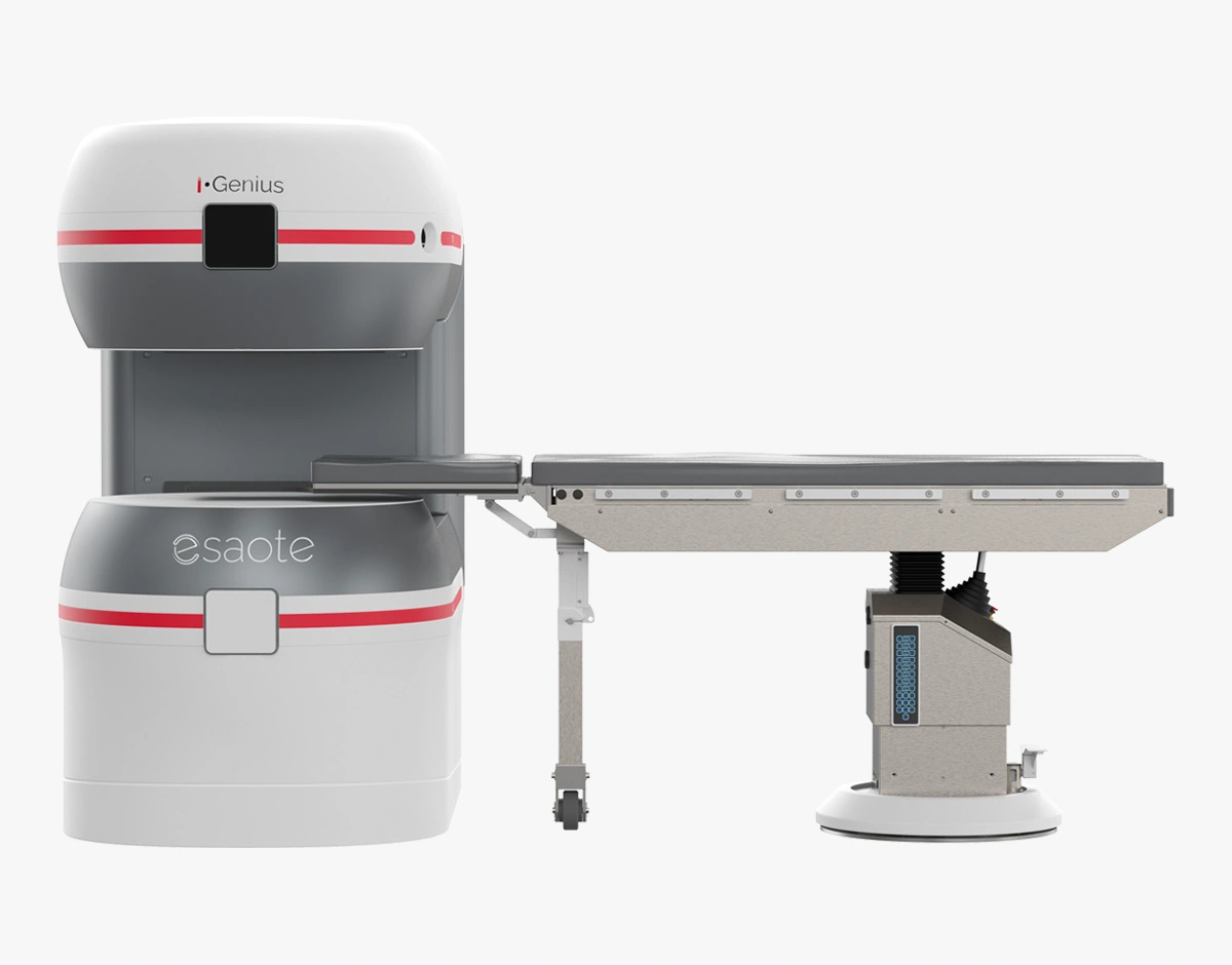
I-Genius
Rezonans śródoperacyjny, który usprawnia przebieg badania dzięki obrazowaniu w czasie rzeczywistym i wspiera neurochirurga w krytycznych decyzjach.
Read more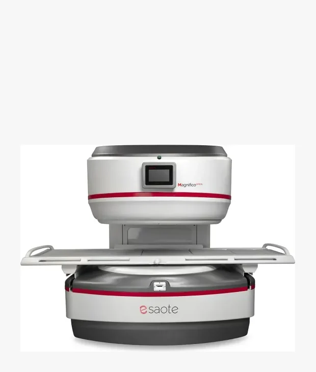
Magnifico Open MRI System
Najnowsza technologia MR dla bezkompromisowej jakości diagnostycznej, w połączeniu z magnesem stałym gwarantującym niskie koszty eksploatacji.
Read more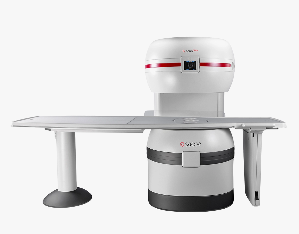
S-scan Open
Połączenie nowatorskiej włoskiej technologii z rezonansem projektowanym pod potrzeby użytkownika — skuteczność diagnostyczną i koszt badania.
Read more